
PRODUCTS SOLD ON PEPTIDESLABUAE.COM ARE FOR RESEARCH PURPOSES ONLY AND ARE NOT FOR HUMAN OR VETERINARY USE.
£79.99
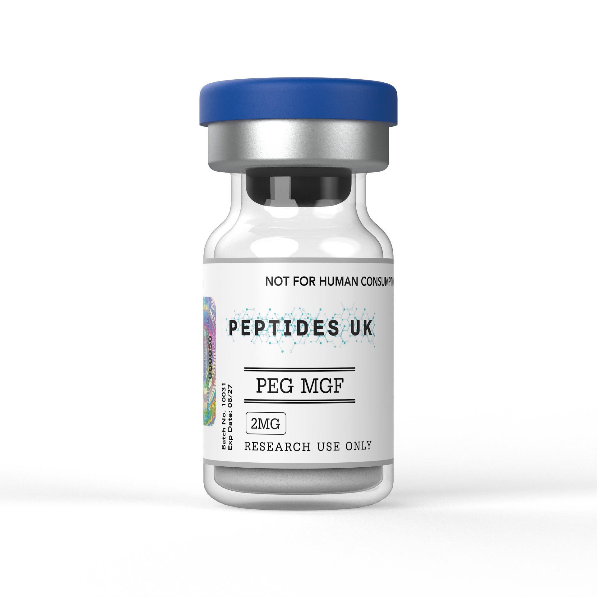
Buy PEG MGF Peptide in UAE – In Stock & Ready to Ship
PEG MGF (PEGylated Mechano Growth Factor) is a widely researched peptide known for its role in muscle repair and extended cellular regeneration studies. Each batch is independently verified at ≥99% purity and comes with a full Certificate of Analysis (COA) and HPLC testing documentation — giving UAE research teams the confidence they need when sourcing peptides for serious work.
For research use only. Not intended for human or veterinary use.
PEG MGF (Pegylated Mechano Growth Factor) is a synthetic, polyethylene glycol-stabilised analogue of MGF — a splice variant of IGF-1 produced locally in response to mechanical stress and muscle damage — and one of the most specifically engineered tissue repair and muscle biology research peptides available to laboratories in the UAE, combining the receptor-binding activity of the IGF-1Ec C-terminal peptide with PEG modification that dramatically extends its circulating stability, making it a key research tool for studying satellite cell activation, muscle repair mechanisms, local IGF-1 splice variant biology, and the cellular response to mechanical loading and tissue injury. Researchers and institutions across the UAE, Dubai, Abu Dhabi, and the wider GCC can source verified, research-grade PEG MGF with fast international dispatch and full batch documentation included.
✅ ≥99% Purity — HPLC & Mass Spectrometry Verified
✅ Batch-Specific Certificate of Analysis (CoA)
✅ Sterile Lyophilised Powder | GMP Manufactured
✅ Fast International Dispatch to UAE & GCC
PEG MGF is the pegylated form of Mechano Growth Factor — a peptide derived from the unique C-terminal extension (E-domain) of the IGF-1Ec splice variant, which is the human form of MGF produced in skeletal muscle, cardiac muscle, bone, and other mechanosensitive tissues in response to mechanical loading, stretch, damage, or hypoxia. MGF was first identified by Professor Geoffrey Goldspink at University College London through research characterising how IGF-1 gene expression in mechanically loaded muscle produces distinct splice variants with different biological roles — establishing that the IGF-1 gene does not simply produce one growth factor but rather a family of related peptides with tissue-specific and stimulus-specific expression patterns that serve distinct biological functions in tissue maintenance and repair.
The native MGF peptide has a very short half-life in biological systems — rapidly degraded by serum proteases within minutes of entering circulation, which reflects its physiological role as a locally acting paracrine and autocrine factor rather than a systemic hormone. While this local, transient activity is appropriate for its endogenous function in responding to acute mechanical stress at the site of tissue loading, it severely limits the research utility of native MGF in pre-clinical models where systemic administration is required and sustained receptor engagement is needed for meaningful experimental outcomes. PEGylation addresses this limitation directly — the attachment of polyethylene glycol chains to the MGF peptide creates a steric shield around protease cleavage sites, dramatically reducing degradation rates and extending circulating half-life from minutes to days while preserving the peptide’s capacity to engage its receptor and activate downstream signalling.
The biological target of MGF’s C-terminal E-domain peptide is distinct from the classical IGF-1 receptor — research has characterised MGF as acting through a receptor system separate from IGF-1R, engaging a binding site that triggers satellite cell activation and proliferation through mechanisms that are complementary to but distinct from IGF-1 receptor signalling. This receptor distinction is biologically important — it means MGF and systemic IGF-1 serve different roles in muscle biology, with MGF acting as the acute local responder to mechanical damage driving satellite cell activation, while systemic IGF-1 mediates the longer-term differentiation and maturation phases of muscle repair and growth.
In laboratory settings, PEG MGF research is centred on its extended-stability MGF receptor engagement and the downstream consequences of satellite cell activation and tissue repair signalling. Research applications include:
Its unique combination of MGF receptor specificity and PEG-extended circulating stability makes PEG MGF the practical research tool of choice for studying MGF biology in systemic pre-clinical models — accessing the satellite cell activation and tissue repair biology of mechanical loading response in experimental designs where native MGF’s minute-long half-life makes it unusable. All applications are for research use only.
PEG MGF and its parent compound MGF have generated a significant and growing research literature centred on satellite cell biology, muscle repair mechanisms, IGF-1 splice variant pharmacology, and the tissue response to mechanical loading and injury.
MGF discovery and splice variant research established the foundational biology underpinning PEG MGF’s research relevance — with Professor Goldspink’s group at UCL characterising how mechanical loading and muscle damage trigger expression of the IGF-1Ec splice variant in skeletal muscle, producing MGF as a local paracrine signal distinct from systemic liver-derived IGF-1. Research established that MGF expression precedes systemic IGF-1 elevation following muscle damage — appearing acutely at the injury site to initiate satellite cell activation before systemic IGF-1 drives the subsequent differentiation and maturation phases. This temporal sequence has been central to understanding the coordinated biology of muscle repair and has positioned MGF as the initiating signal in the satellite cell activation cascade.
Satellite cell biology research has characterised MGF’s effects on muscle stem cells — with studies documenting MGF-driven satellite cell activation from quiescence, enhanced proliferation of myoblasts, and increased muscle precursor cell numbers in response to MGF signalling. Research has established that MGF’s C-terminal E-domain peptide activates satellite cells through a receptor mechanism distinct from IGF-1R — providing mechanistic separation between MGF-driven satellite cell activation and IGF-1-driven satellite cell differentiation, and establishing the two signals as sequential and complementary phases of the muscle repair response rather than redundant pathways.
PEGylation pharmacokinetic research has directly characterised the stability advantage conferred by PEG modification — with studies comparing native MGF and PEG MGF half-lives in serum and documenting the dramatically extended circulating stability of the pegylated form. Research has confirmed that PEGylated MGF retains the biological activity of native MGF at its receptor while achieving systemic distribution and prolonged receptor engagement that native MGF cannot provide — validating PEGylation as the appropriate modification strategy for translating MGF biology from local paracrine research into systemic pre-clinical research models.
Cardiac muscle research has examined MGF expression in the heart — with studies documenting MGF production in cardiac muscle in response to mechanical stress and ischaemic injury, and characterising MGF-driven cardiomyocyte protection and repair responses. Research examining PEG MGF in cardiac ischaemia models has reported cardioprotective effects associated with MGF receptor activation in cardiac tissue — establishing cardiac biology as a secondary but significant research area alongside the primary skeletal muscle focus.
Neuroprotection research has identified MGF receptor expression in neuronal tissue and characterised neuroprotective effects associated with MGF signalling — with studies reporting reduced neuronal apoptosis and improved survival parameters in injury models treated with MGF, expanding the research relevance of PEG MGF into CNS biology beyond its primary musculoskeletal research context.
Age-related muscle biology research has examined MGF expression decline with ageing — with studies documenting reduced MGF splice variant production in aged muscle in response to mechanical loading compared to young muscle, providing a mechanistic basis for age-associated impairments in muscle repair capacity and positioning MGF biology as relevant to sarcopenia and age-related muscle loss research.
| Compound | Type | Mechanism | Half-Life | Primary Research Focus | Research Profile |
|---|---|---|---|---|---|
| PEG MGF | Pegylated MGF C-terminal peptide | MGF receptor — satellite cell activation | Days (PEG-extended) | Muscle repair, satellite cell biology | Well-documented |
| Native MGF | MGF C-terminal E-domain peptide | MGF receptor — local paracrine | Minutes | Reference MGF biology, local studies | Well-documented |
| IGF-1 LR3 | Long-acting IGF-1 analogue | IGF-1R — systemic anabolic | ~20 hours | IGF-1 axis, anabolic biology | Extensively studied |
| IGF-1 DES | Truncated IGF-1 analogue | IGF-1R — local tissue activity | Short | Local IGF-1 biology, tissue repair | Well-documented |
| Systemic IGF-1 | Endogenous growth factor | IGF-1R — full agonist | ~12–15 hours | Reference IGF-1 biology | Extensively studied |
| TB-500 (Thymosin β4) | Actin-sequestering peptide | Actin dynamics, tissue repair | Extended | Tissue repair, regeneration | Well-documented |
| BPC-157 | Gastric pentadecapeptide | Growth factor signalling | Extended | Tissue repair, cytoprotection | Well-documented |
| Parameter | Detail |
|---|---|
| Type | Pegylated IGF-1 Splice Variant C-Terminal Peptide |
| Parent Compound | Mechano Growth Factor (MGF) — IGF-1Ec E-domain |
| Modification | Polyethylene glycol (PEG) — protease resistance and half-life extension |
| Molecular Weight | ~2867 Da (peptide) + PEG modification |
| Mechanism | MGF receptor engagement — satellite cell activation |
| Key Advantage | Days-long stability vs minutes for native MGF |
| Purity | ≥99% |
| Verification | HPLC & Mass Spectrometry |
| Form | Lyophilised Powder |
| Solubility | Sterile water or suitable laboratory buffer |
| Storage | -20°C, protected from light and moisture |
| Intended Use | Research use only |
Every order dispatched to the UAE and GCC includes:
Yes. We supply research-grade PEG MGF with international dispatch to the UAE, Dubai, Abu Dhabi, Sharjah and across the GCC. All orders include full batch documentation and are packaged to maintain peptide integrity throughout transit. This compound is supplied strictly for laboratory research use only.
Mechano Growth Factor is a splice variant of the IGF-1 gene — produced from the same gene as systemic IGF-1 but through alternative mRNA splicing that incorporates a unique C-terminal E-domain sequence (the Ec extension) not present in liver-derived systemic IGF-1. While systemic IGF-1 is produced primarily in the liver in response to growth hormone and acts as a circulating endocrine growth factor engaging IGF-1R across the body, MGF is produced locally in mechanosensitive tissues — particularly skeletal and cardiac muscle — in response to mechanical loading, damage, or hypoxia, acting as a local paracrine signal through a receptor distinct from IGF-1R. The biological distinction is significant — MGF drives the initial acute satellite cell activation response to muscle damage, while systemic IGF-1 mediates the subsequent differentiation and maturation phases. The two molecules are complementary regulators of tissue repair biology rather than redundant versions of the same signal.
Satellite cells are muscle stem cells that reside in a quiescent state between the sarcolemma and basal lamina of mature muscle fibres — serving as the primary regenerative reserve of adult skeletal muscle. Following mechanical damage or intense loading, satellite cells are activated from quiescence, proliferate to generate a pool of myoblasts, and subsequently differentiate and fuse to repair damaged fibres or form new muscle tissue. MGF has been identified as one of the primary signals driving satellite cell activation from quiescence — the initial and rate-limiting step in the muscle repair cascade. Understanding the molecular mechanisms through which MGF activates satellite cells has broad implications for muscle biology research, age-related muscle loss research, and the biology of tissue regeneration — making satellite cell activation the central research question in the MGF and PEG MGF literature.
Native MGF is designed by biology to act as a local, transient paracrine signal — produced at the site of mechanical stress, acting briefly on nearby satellite cells, and then rapidly degraded. This local, short-lived activity is biologically appropriate for its endogenous function but makes native MGF practically unusable in systemic pre-clinical research models where it is degraded within minutes of administration before reaching target tissues in meaningful concentrations. PEGylation — the attachment of polyethylene glycol chains to the peptide — creates steric shielding around protease cleavage sites, dramatically reducing degradation and extending circulating half-life to days. This transforms MGF from a locally acting paracrine factor into a systemically distributable research compound capable of reaching target tissues and maintaining receptor engagement across experimental protocol timeframes — making PEGylation not merely a refinement but a prerequisite for meaningful systemic pre-clinical MGF research.
PEG MGF and systemic IGF-1 act through distinct receptor systems and serve sequential roles in the tissue repair cascade — making their interaction in research models a question of complementary rather than competing biology. Research examining combined MGF and IGF-1 axis signalling has characterised how MGF-driven satellite cell activation creates the proliferating myoblast pool that IGF-1 receptor signalling subsequently differentiates into mature muscle tissue. In pre-clinical research models, PEG MGF’s extended stability allows study of how sustained MGF receptor engagement influences the subsequent IGF-1 axis-dependent phases of muscle repair — providing insights into the temporal coordination between local MGF signalling and systemic IGF-1 biology that would be impossible to examine with native MGF’s minute-long half-life.
Yes — while skeletal muscle is the primary and most extensively studied tissue in MGF research, MGF expression and biological activity have been characterised in several other mechanosensitive tissues. Cardiac muscle research has documented MGF production in response to mechanical stress and ischaemic injury, with studies characterising cardiomyocyte protection and repair responses associated with MGF receptor activation. Bone biology research has examined MGF expression in osteoblasts and mechanically loaded bone tissue. Neuronal research has identified MGF receptor expression in the CNS and characterised neuroprotective effects in injury models. These findings have expanded MGF’s research relevance beyond its original skeletal muscle context — establishing it as a broader mechano-responsive tissue repair signal with biological roles across multiple tissue types, each representing a distinct and active research area for PEG MGF investigation.
Allow the vial to reach room temperature before opening. Add sterile water or appropriate laboratory buffer slowly down the vial wall and swirl gently without shaking. Prepare at your protocol’s required concentration. Aliquot and store at -80°C to minimise freeze-thaw degradation and maintain peptide integrity between experimental sessions. Avoid repeated freeze-thaw cycles and exposure to elevated temperatures. Handle with standard peptide protocols to preserve the integrity of the PEG modification and biological activity.
Orders are dispatched promptly via tracked international courier. Delivery to the UAE typically takes 3–5 working days, with packaging designed to maintain peptide stability and integrity throughout transit.
PEG MGF is supplied exclusively for legitimate scientific research conducted within licensed laboratory environments. This product is not intended for human consumption, self-administration, or any therapeutic or veterinary application. It must be handled solely by qualified researchers in compliance with applicable UAE regulations and institutional ethics guidelines. By purchasing, you confirm this compound will be used exclusively for approved in vitro or pre-clinical research purposes.

WhatsApp us
Receive News